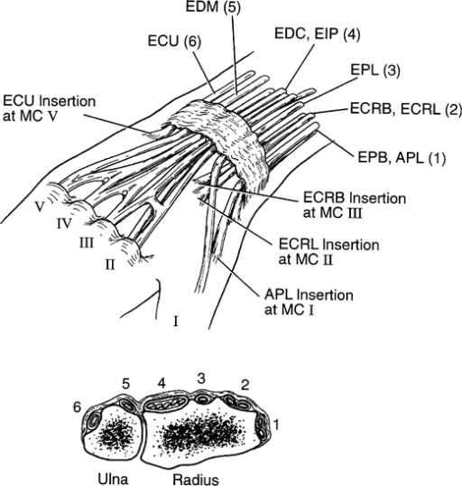

The present study aims to demonstrate the microvascular supply to this composite flap in a cadaveric model.
EXTENSOR COMPARTMENTS SKIN
This composite tissue flap would provide vascularised tendon graft, the overlying skin and a potential intervening gliding plane, thus enhancing tendon excursion. We postulate the use of a vascularised composite flap of extensor retinaculum and the overlying skin to reconstruct complex dorsal digital injuries. However, due to poor tendon vascularity, a scarred tissue bed or underlying fractures, adhesion formation often prevails.

In delayed operations, extensor tenolysis may be used to resolve adhesions interrupting tendon excursion. In acute injuries, either staged reconstruction or the use of a non-vascularised tendon graft can be considered. While reconstruction of individual anatomical components is feasible, re-establishing these areolar gliding planes for tendon excursion remains a surgical challenge. Interruption of this gliding plane results in reduced range of movement, joint contracture, pain and functional loss in the affected digit. It has a broad gliding interface with its overlying subcutaneous areolar tissue and the underlying metacarpal or phalangeal periosteum.

1,2 The extensor mechanism consists of a flat, delicate and complex tendon system. Keywords: tendons, tendon injuries, surgical flapsĬomplex extensor tendon injuries of the fingers can be associated with a slow and unpredictable recovery. It can be employed for simultaneous vascularised tendon and skin reconstruction. The skin overlying the extensor retinaculum was predictably supplied by either artery through the perforator vessels between the fourth and fifth extensor tendon compartments.Ĭonclusion: A composite unit of skin and extensor retinaculum can be harvested on either the anterior or posterior interosseous arteries. Results: The anterior and posterior interosseous arteries supplied the extensor retinaculum through a dense network of vessels with choke anastomoses. Specimens were subsequently assessed by digital subtraction angiography to evaluate the corresponding microvascular supply to the composite flap. Specimens were then dissected to raise the proposed composite flap of extensor retinaculum and the overlying integument. The anterior (n=9) or posterior (n=9) interosseous artery was exposed and selectively injected with a coloured dye. Methods: An anatomical study of 18 cadaveric upper limbs was conducted to investigate the technical feasibility of a composite flap prior to its clinical application. This anatomical study evaluates vascular supply to a suitable composite flap comprising skin, subcutaneous tissue and extensor retinaculum. Vascularised tissue to reconstruct the skin and extensor defect would be the ideal reconstruction in both the acute and delayed settings. This is an open access article distributed under the Creative Commons Attribution Licence which permits unrestricted use, distribution and reproduction in any medium, provided the original work is properly cited.īackground: Complex digital extensor tendon injuries are difficult to manage when adhesion formation and stiffness prevail. Authors retain their copyright in the article. The composite extensor retinaculum cutaneous flap: an anatomical cadaveric study. William A Cuellar BAppSc, 1 Siddharth Karanth MBBS 1,2ĭepartment of Plastic and Reconstructive SurgeryĪddress: Department of Plastic and Reconstructive SurgeryĬitation: Sreedharan S, Ross RJ, Froelich JJ, Cuellar WA, Karanth S. The composite extensor retinaculum cutaneous flap: an anatomical cadaveric study Sadhishaan Sreedharan MBBS, 1,2 Richard J Ross MBBS PhD, 2 Jens J Froelich MD PhD, 1,3 The composite extensor retinaculum cutaneous flap: an anatomical cadaveric study


 0 kommentar(er)
